“What you can’t see, doesn’t exist!” – this saying has its place when it comes to dentistry.
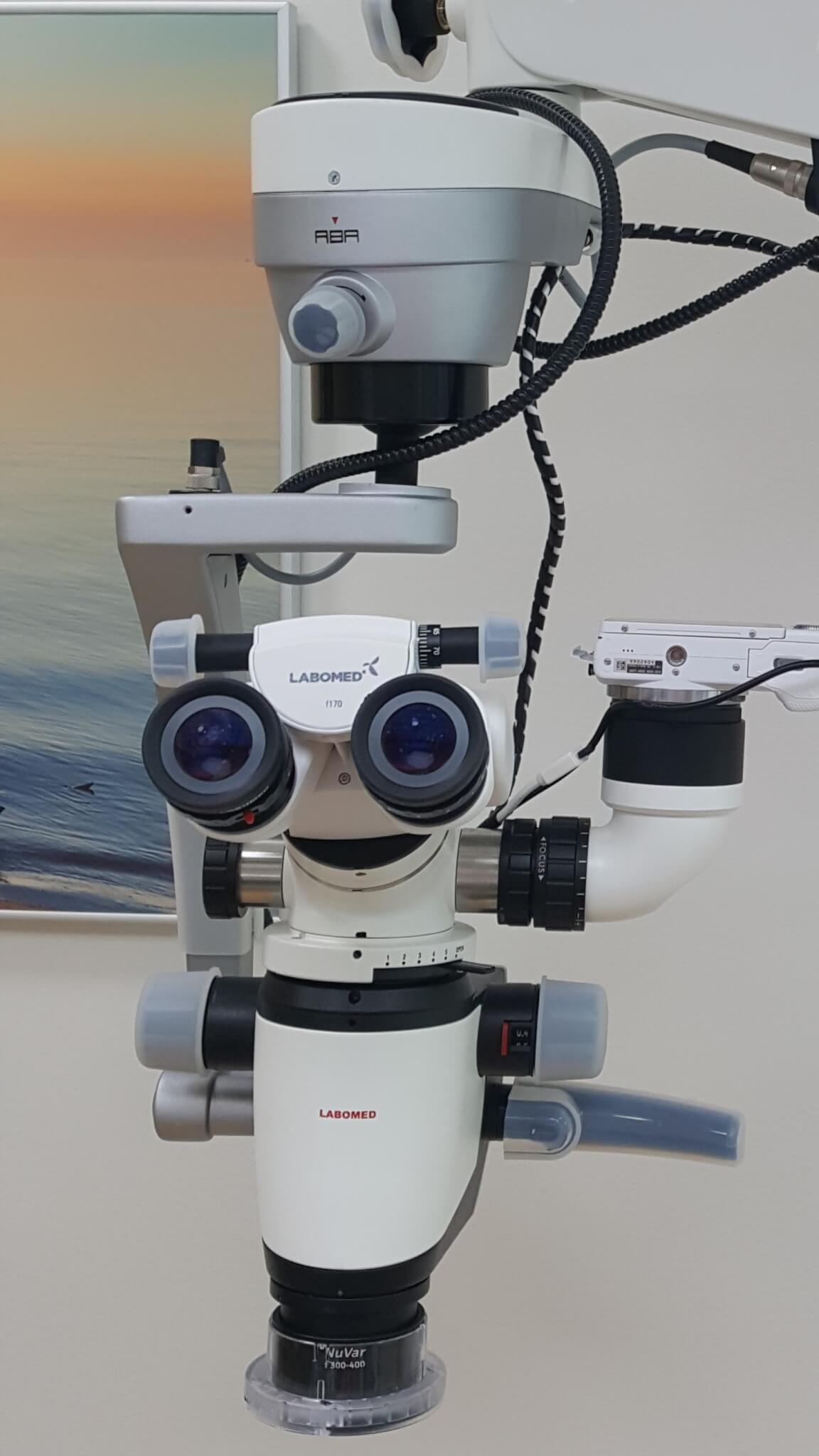
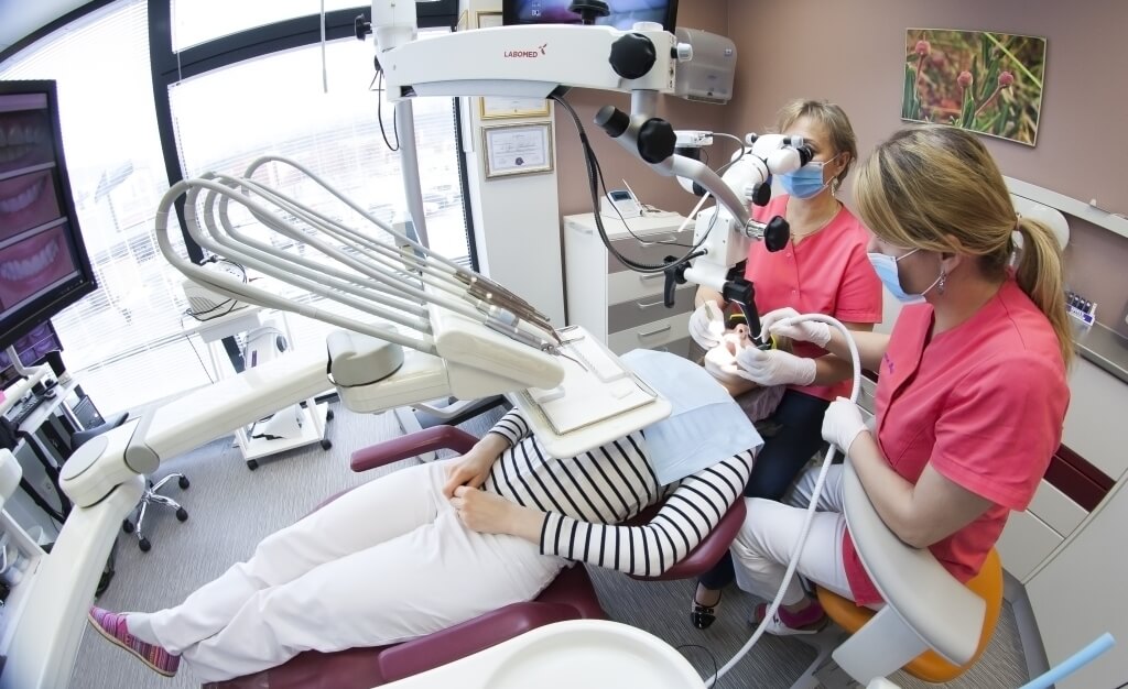
In modern dentistry, a microscope is both an assistant, a controller and a diagnostician. We use a microscope for caries treatment, root canal treatment, prosthetic and surgical procedures. Also in a situation where what you see with your eyes raises questions, and to answer these, you just need to SEE more. Using a microscope allows you to observe all the small details, cracks and nooks.
For example, root canal treatment is difficult and time-consuming, especially for multi-rooted teeth located at the back of the jaw. The microscope magnifies the field of action and also directs powerful light there. Root canal treatment with a microscope helps identify and handle even the smallest important details.
Dental treatment with a microscope allows for a better quality result thanks to accuracy, clarity and certainty in making treatment decisions.
The fast pace of life forces us to rush. We want to get everything now and quickly. You can’t rush when working with a microscope, but the microscope increases the quality of the entire work process saving us treatment time and costs.
“Rush slowly” is a fitting saying in this situation.
But more importantly – the lifetime of the treated tooth increases.
As does the joy you get from the work well done!
Microscope is connected with imaging and video technology, which can be used to record work processes and explain them later.
But often “words are not needed”, because what you see with your eyes speaks for itself.