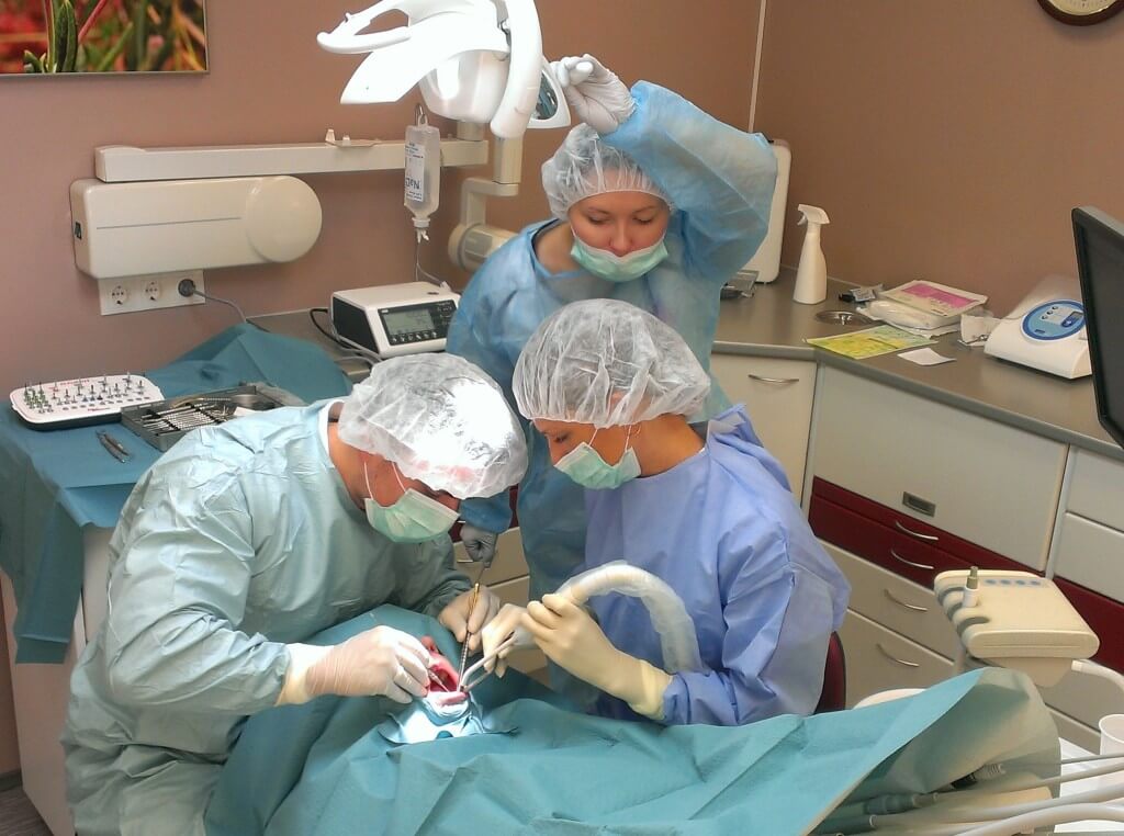
We offer the following oral and dental surgery services:
Ridge augmentation
When a tooth is removed and the support from the root is lost, the jawbone surrounding it begins to shrink. The bone gradually deteriorates, as a result of which the alveolar ridge becomes thinner and more narrow.
To restore the masticatory function with dental implants, a sufficient amount of bone mass is needed to support the implant.
There are several options for bone grafting or more specifically for ridge augmentation:
- Autogenous bone block (patient’s own bone) harvesting from another part of the mouth or body and its transfer to the deteriorated area;
- Collection of autogenous bone by scraping or drilling another part of the mouth or body and its transfer to the deteriorated area;
- Filling the deteriorated area with artificial bone.
Using autogenous bone is quite uncomfortable for the patient and the recovery period post-surgery is longer and more difficult.
However, the use of artificial bone is not as effective as the use of autogenous bone.
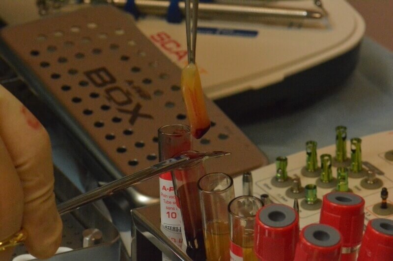
The use of A-PRF and I-PRF mixed with synthetic bone granules allows us to avoid the difficult process of harvesting autogenous bone while providing the same results as autogenous bone.
The growth factors in A-PRF and I-PRF stimulate bone growth around artificial bone granules and together they form the new bone structure on which the new implant can be placed.
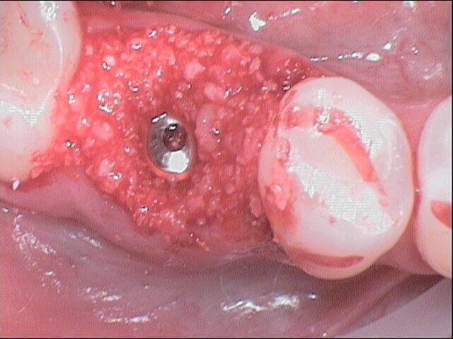
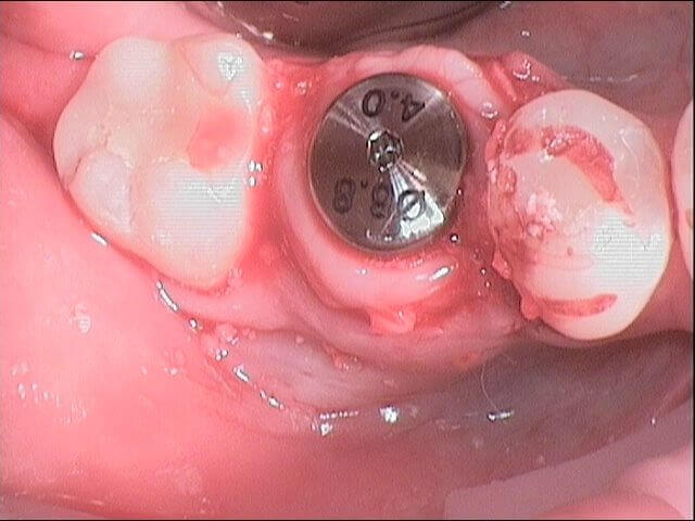
We always use A-PRF and I-PRF extracted from the patient’s own blood in ridge augmentation. The implant can be placed approximately 3–4 months after ridge augmentation.
Sinus lift
When an upper tooth is removed, the surrounding bone starts to deteriorate quite quickly and, as a result, the layer of bone separating the oral cavity and the maxillary sinus becomes so thin that the placement of a dental implant is impossible without bone grafting.
This type of bone grafting is called maxillary sinus floor augmentation or sinus lift.
There are two ways to perform a sinus lift:
- the lateral approach – a hole is drilled in the lateral wall through which the bone grafting is carried out;
- the crestal approach – the bone-grafting material is inserted into the sinus cavity through the drilled implant hole.
The bone growth and healing is significantly faster if the synthetic bone granules have been mixed with A-PRF.
To do this, venous blood is collected from the patient before sinus lift surgery and, after the centrifugation of their blood, A-PRF is obtained.
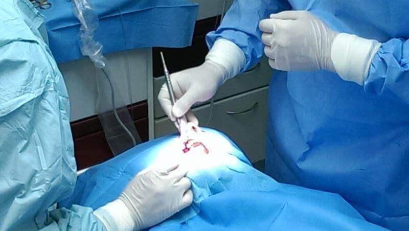
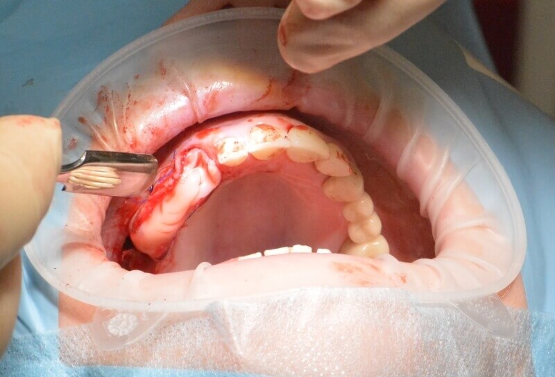
Soft tissue surgery
Studies have shown that thin gums increase the risk of bone loss. However, when it comes to thick gums with better blood supply, the risk of bone loss is significantly smaller. Bone loss causes the neck of the teeth to become exposed.
In addition, gums must be thick enough to support the crowns on implants and to reduce bone loss around the implant.
In the case of extensive ridge augmentation, there is usually not enough gum tissue to cover the increased bone volume. More gum tissue needs to be grown.
There are different ways to grow gum tissue. One method is gingival grafting, which involves harvesting gum tissue from another area in the mouth, e.g. the roof, and then relocating it to the necessary place. It is quite uncomfortable for the patient and causes pain for some time after surgery.
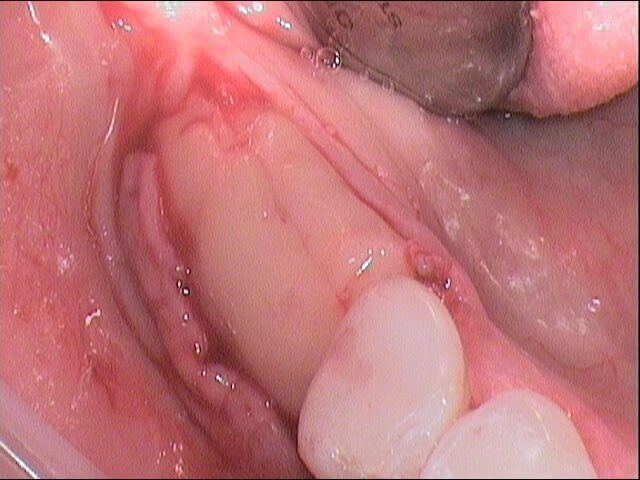
A more comfortable method for the patient is a technique whereby an A-PRF clot prepared from their own blood by centrifugation is attached to the alveolar ridge or the exposed tooth neck. After 2–4 weeks the A-PRF clot has formed a healthy, pink gum tissue.
Thin gums also cause bone loss which in turn causes the loss of interdental papilla which results in black triangles between the teeth. Papilla can be restored and grown by injecting I-PRF into them.
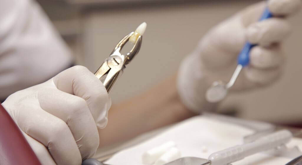
Tooth extraction
The most common causes of tooth extraction are as follows:
- extensive dental caries and resulting inflammation that does not respond to treatment;
- advanced periodontal diseases;
- impacted teeth;
- erupted wisdom teeth that have no room to grow or are decayed and their treatment is not practical;
- fractured tooth root or crown.
Before tooth removal local anaesthesia is used to numb the area, thus the removal of a noninflammatory tooth is usually painless.
After extraction
After the extraction a blood clot forms in the tooth socket to protect the wound. Soon after, white granulation tissue will cover the wound and over time will turn into connective tissue. The wound will heal within 5–10 days but it will take months for the bone tissue to fully heal.
We use A-PRF clots placed in the alveolar socket to promote the healing of bone tissue. The A-PRF clot prevents the collapse of the alveolar ridge and boosts the growth of new bone tissue.
Placing the A-PRF in the alveolar socket can prevent the need for ridge augmentation later on, and after 2–4 months, it is possible to place an implant over the place of the removed tooth. Immediate implantation after tooth extraction is also possible by filling the empty alveolar socket around the implant with a mixture of A-PRF and artificial bone.
In both cases, A-PRF plays an important role in bone recovery and healing.
It is not advisable to eat or drink for a few hours after tooth extraction. Smoking is also not allowed. Care should be taken when brushing the teeth to avoid damaging the blood clot in the socket. Starting the day after surgery, chlorhexidine solution should be used to rinse the mouth for a week. The solution is available to purchase at our clinic and in pharmacies. A salt water rinse may also be used.
If you have pain in the days following the tooth extraction, you should return to the clinic. If necessary, the doctor will clean the wound and prescribe anti-inflammatory treatment.
Soft tissue laser surgery
A laser can be used for crown lengthening by recontouring gum tissue. Gingival enlargements and small fibrous epulides can also be treated or removed with a laser.
Lasers have an antimicrobial effect and boost cellular resistance.
Lasers can be used to reshape the gum line before taking an impression for prosthesis. Lasers are essential for exposing wisdom teeth that have only partially erupted.
We use LaserSmile 810 nm diode laser.