We need information to prepare a comprehensive treatment plan and to make the right treatment decisions.
We use a variety of devices and tools for diagnosis:
- 3D X-ray
- Intraoral X-ray
- Intraoral video camera
- Microscope
- Intraoral 3D scanner
- Shade-matching device
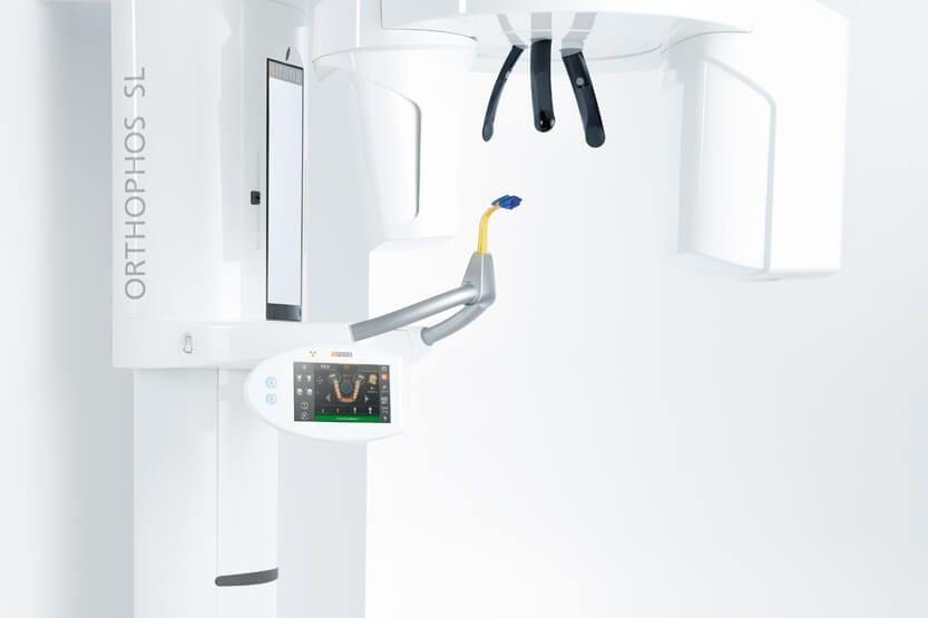
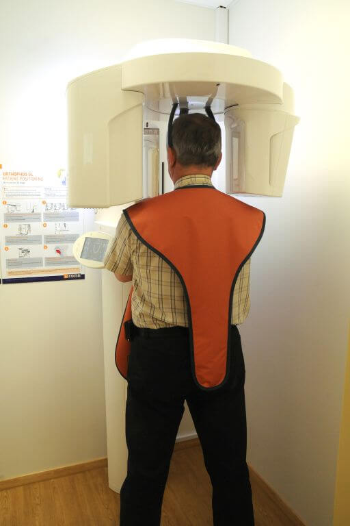
3D X-ray
A 3D X-ray is essential for assessing the condition of the jawbone and tooth roots. We use the Sirona Orthophos SL 3D. This device allows us to take both 3D X-ray and panoramic X-ray images. We can discover and identify previously unseen changes beneath the teeth and in bone structure, discuss the possibility of wisdom tooth extraction, prepare the placement of implants with the help of special software, detect the presence of tumours and cysts, and assess the possibility of surgical and endodontic treatment.
Taking an X-ray image with the new 3D device is quick and the patient radiation dose is minimal. We use a special X-ray apron to protect the patient. We have a separate room for taking 3D X-rays which has radiation shielded walls that meet modern requirements.
X-ray images are stored in a database where the doctor can access them quickly at any time.
Lumen Dental Clinic offers 3D and panoramic X-rays as a separate service. The patient receives the image file on a CD as well as the software for viewing it.
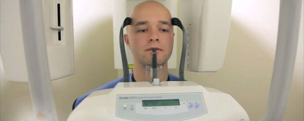
Intraoral X-ray
Intraoral X-ray is necessary to quickly assess the condition of 1–3 teeth. Intraoral X-rays can also be used to detect root canal infections and hidden caries. Additionally, it is essential for assessing the condition of the root canal during root canal treatment, and for taking check-up images after the placement of crowns, implants and implant crowns.
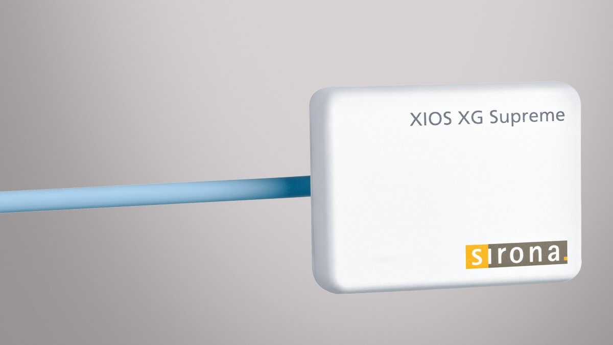
We use the Sirona XIOS XG Supreme intraoral sensor and Carestream X-ray imaging system for taking intraoral X-ray images.
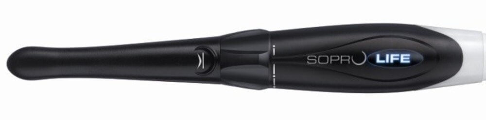
Intraoral video camera
Using the intraoral camera allows both the doctor and the patient to see the condition of the patient’s mouth and teeth on a big screen. During the appointment, the doctor can quickly take photos of the problematic teeth and the whole mouth to analyse them later when preparing the treatment plan. The camera allows the doctor to view the teeth at an angle that would be difficult to see with traditional examination methods. The high-resolution camera sensor shows big close-up images on the screen which can also be shown to the patient.
Intraoral cameras are a practical tool that can be used to visually explain the nature of the problem to the patient and to educate them about it.
We use Lensiora and Acteon Sopro cameras.
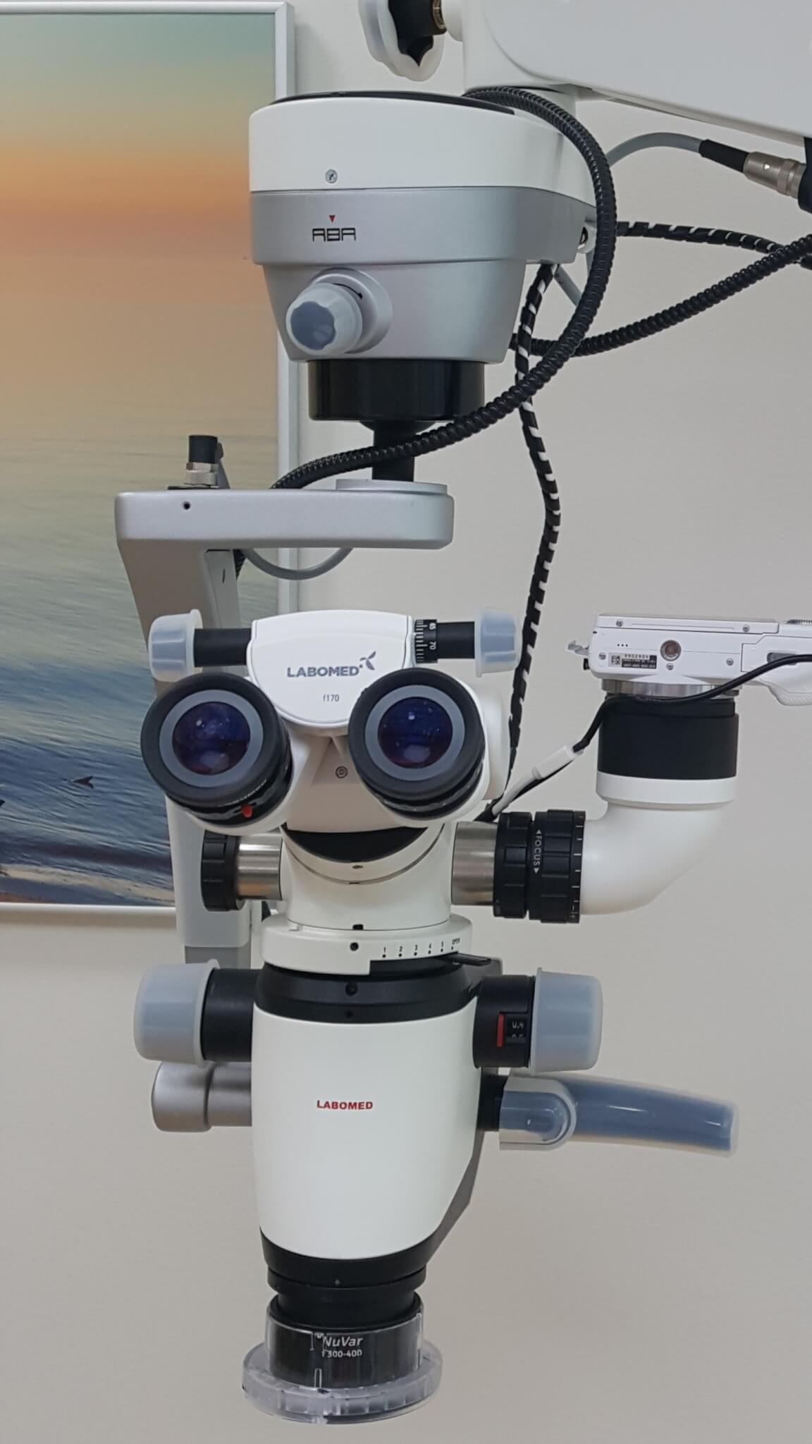
Microscope
Microscopes enable the dentist to see details that are not visible to the naked eye or even with a dental loupe. Microscopic tooth fractures can be detected and treated before they even become a problem. Microscopes enable the detection and diagnosis of dental diseases that would otherwise go undetected.
We use CJ-Optic ja Labomed microscopes.
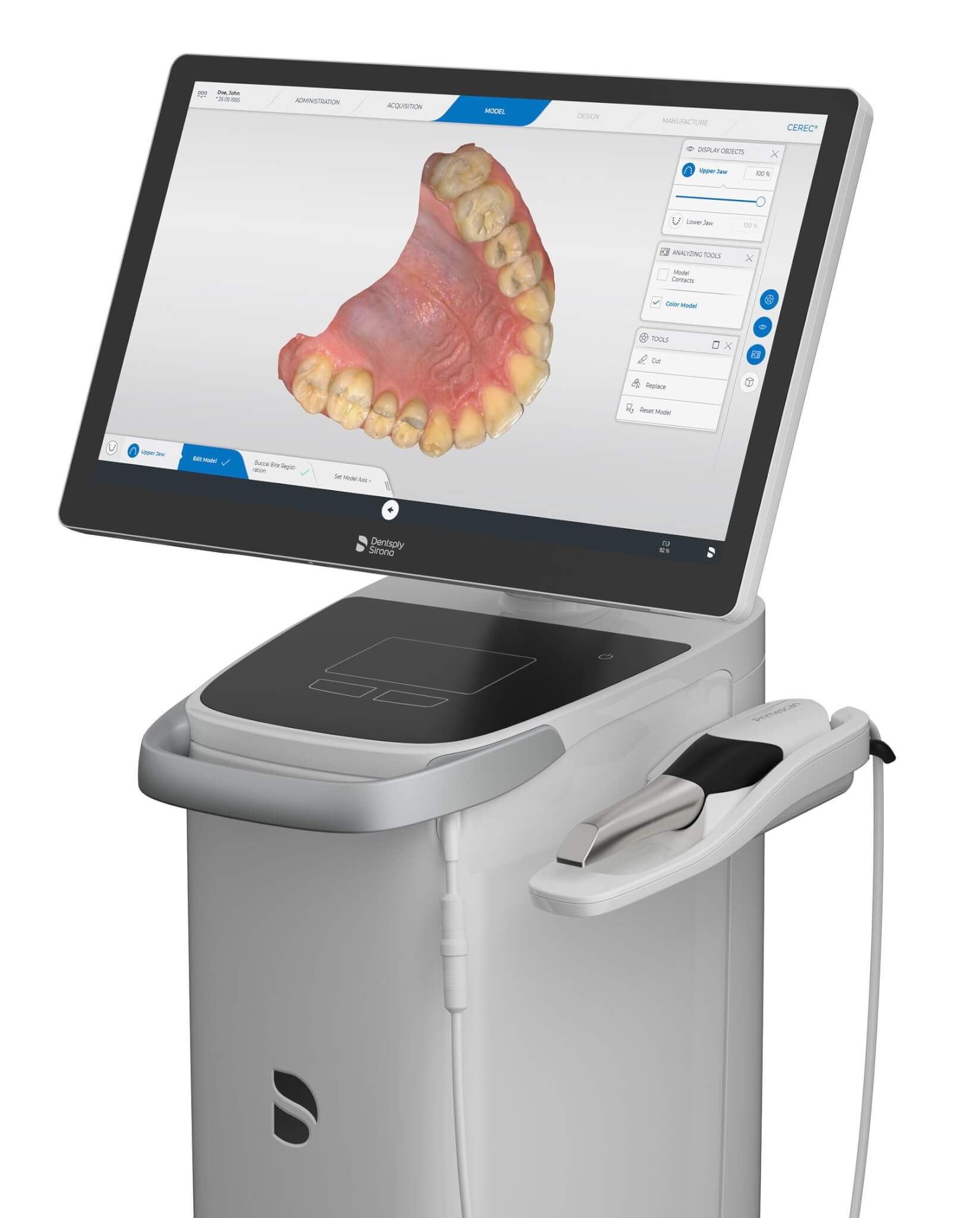
Intraoral 3D scanner
The intraoral 3D scanner provides and opportunity to inspect and analyse the dentition spatially and functionally. Many doctors have used and still continue to use gypsum models to analyse dentition. A digital 3D model provides many more opportunities for inspecting and analysing the current condition and what the objective of the treatment is. The option to view the model from different angles, in part or as a cross-section, to assess dentition and the contact strength of teeth, to analyse jaw movement in the virtual articulator – all of this provides valuable information to the doctors which allows them to create a more accurate treatment plan.
A 3D scan of the mouth is a source of information that can be used in combination with other diagnostic devices for digital treatment planning. For instance, for the placement of implants of root canal treatment.
We use Sirona Cerec Primescan for 3D scanning.
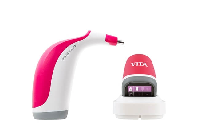
Shade-matching device
In prosthodontics, it is very important to match the colour of the dental crown, bridge or removable denture to the rest of the teeth. As is well-known, our colour perception is very different and highly dependent on the surrounding environment and light. To determine the colour of the tooth without any interferences (e.g. patient’s red shirt) and independently from the doctor’s own colour perception, we use the VITA EasyShade V digital shade-matching device.